
Before pregnancy
Women
Menstrual cycle
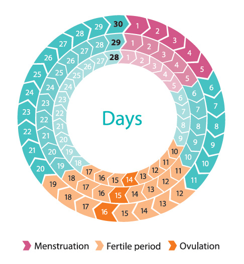
Illustration: Les publications du Québec
Your body prepares for pregnancy during every menstrual cycle.
Menstruation is a stage of the menstrual cycle. Menstrual cycles begin at puberty around the age of 12 and continue until menopause, which typically occurs around age 51.
To determine the length of your menstrual cycle, count the number of days from the beginning of your period to the day before your next period starts. Menstrual cycles can last anywhere from 21 to 35 days, but are usually between 28 and 30 days long.
During a menstrual cycle, your body goes through a number of changes. Many interactions take place between your brain and your pituitary gland, a hormone-secreting organ. These interactions trigger the release of hormones that stimulate ovulation, which in turn prepares your body for fertilization.
Ovulation
Female reproductive system
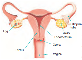
Illustration: Bertrand Lachance
Women are born with all the eggs they will ever have. They have about 400,000 eggs at puberty, and by menopause, all of them are gone.
Ovulation occurs when an ovary releases an egg. Once an egg is released, it is drawn into the fallopian tube. It may then come into contact with sperm, at which point fertilization may occur (see Fertilization).
To estimate when you will ovulate, count backwards 14 days from the end of your menstrual cycle. Women with regular 28-day cycles usually ovulate around the 14th day of their cycle. For women with irregular cycles, however, it is more difficult to predict the day or period of ovulation.
Ovulation period (or fertile period)
Since ovulation does not always occur on the expected day, we use the term ovulation period or fertile period. This is when a woman is most likely to ovulate. If a man and a woman have intercourse during the fertile period, there is a one in four chance (at age 20) and a one in twenty chance (at age 40) that fertilization will occur.
An egg must be fertilized within 12 hours. If it doesn’t come in contact with a sperm during this period, it disappears through vaginal discharge. At this point, the glands in the brain will stop producing hormones. This triggers menstruation, and the cycle starts all over again.
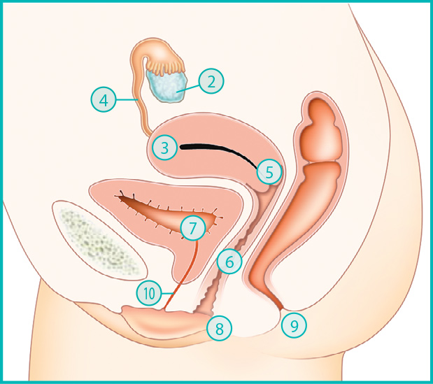
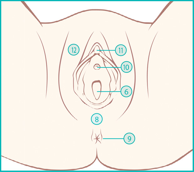
Female anatomy
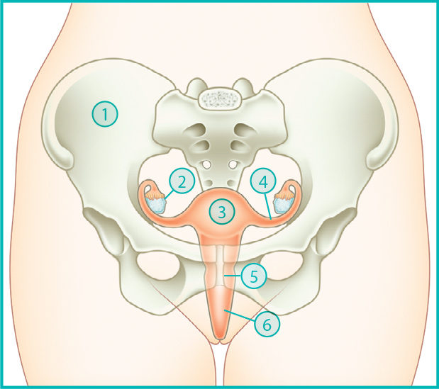
- Pelvis: Bone that supports the organs in the mother’s abdomen.
- Ovary: The two ovaries produce eggs and female hormones.
- Uterus: Muscular organ the size of a small pear that grows as the pregnancy progresses. This is where the embryo develops.
- Fallopian tube: The Fallopian tubes connect the uterus and the ovaries. They transport eggs and are necessary for fertilization.
- Cervix: Bottom part of the uterus connected to the vagina. During menstruation, blood flows from the cervix, which is almost closed. During labour, the cervix dilates to let the baby through.
- Vagina: A roughly 8 cm long passageway between the uterus and the vulva. The vagina is flexible and elastic so it can stretch during intercourse and delivery.
- Bladder: Organ that holds the urine produced by the kidneys.
- Perineum: Viewed from the exterior, the region between the anus and the vulva. The muscles of the perineum form a sort of internal “hammock” that supports the genital organs and bladder.
- Anus: Opening through which feces are expelled.
- Urethra: Tube that carries urine from the bladder to the outside of the body. It is part of the perineum.
- Clitoris: Sensitive, erogenous organ that plays an important role in female sexual pleasure.
- Vulva: All external genitalia, including the labia and clitoris.
Men
Throughout their lives, men produce sperm. Sperm production begins at puberty and continues until death.
Sperm are produced in the testicles (see below Male anatomy), where they go through a number of stages. It takes about two and a half months before they are ready for fertilization. Once they are ready, they are stored in the seminal vesicles.
When a man ejaculates, sperm from the seminal vesicles are mixed with fluids from the prostate and other glands of the male reproductive organs. This is known as semen.
The semen from a single ejaculation usually contains between 20 million and 200 million sperm cells. Sperm can live 72 to 120 hours in a woman’s genital tract, but only a few seconds outside it.
Male anatomy
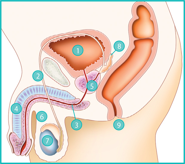
- Bladder: Organ that holds the urine produced by the kidneys.
- Vas deferens: Tube that carries sperm from the testicles to the prostate.
- Urethra: Tube that carries urine from the bladder and out the penis. It also carries semen from the prostate and out the penis.
- Penis: Male genital organ. Its sponge-like tissue swells with blood during erections.
- Prostate: Gland that secretes seminal fluid, one of the substances composing semen.
- Scrotum: Sac of skin that protects the testicles.
- Testicle: The testicles (or testes) are the organs that produce sperm.
- Seminal vesicle: Located above the prostate, the seminal vesicles are reservoirs that store sperm that are ready for fertilization.
- Anus: Opening through which feces are expelled.
Illustrations: The Pregnancy Book
Adapted by Bertrand Lachance
with the permission of the Department of Health, UK
Egg: Reproductive cell produced by the ovary. When an egg and a sperm fuse, an embryo may form.
Embryo: Name given during the first full 10 weeks of pregnancy to the human being developing in the mother’s abdomen.
Fertilization: Fusion of a sperm and egg.
Labour: Process by which the baby passes from the uterus to the outside world, primarily through contractions of the uterus.
Sperm: Reproductive cell produced in the testicles. When a sperm and egg fuse, an embryo may form.